
 Gold Member
 Business License
X-ray diffraction technology - seeing through the order of the crystal world in materials
Abstract: X-ray diffraction technology is a core analysis method that uses the interaction between X-rays and crystalline materials to produce diffraction effects to analyze the microstructure of materials. Since its discovery by Laue and others in 1912, this technology has become an indispensable cornerstone in the fields of materials science, chemistry, physics, biology and geology. This article aims to systematically explain the basic principles of X-ray diffraction, main technical methods (such as single crystal and polycrystal diffraction), its application in structural analysis and phase analysis, and explore its latest progress in cutting-edge directions such as high-throughput, in-situ and micro-area analysis.
Technology development history
The development of XRD technology is closely related to breakthroughs in basic physics research, and its key milestones have established the theoretical and technical framework of modern diffraction analysis.
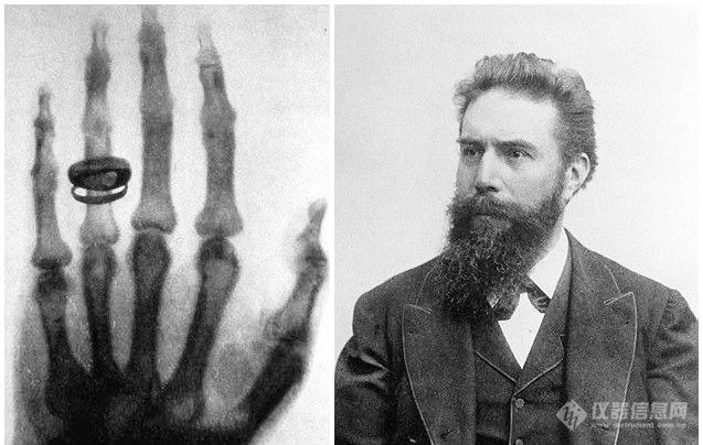
When Roentgen discovered X-rays, he took a photo of his wife's right hand with a ring (picture from the Internet)
1. Foundational stage (late 19th to early 20th century)
Roentgen's discovery of X-rays in 1895 laid the foundation for the birth of diffraction technology. From 1908 to 1909, Walter and Pohl observed the interference fringes that appeared after X-rays passed through a pointed aperture, which provided early clues for the confirmation of the diffraction phenomenon. In 1912, Laue experimentally confirmed the diffraction phenomenon when X-rays meet crystals. This not only proved the electromagnetic wave properties of X-rays, but also created the emerging discipline of X-ray diffractology and became the first milestone in this field.
2. Theoretical and technical forming stage (early to mid-20th century)
In 1913, W.L. Bragg (William Lawrence Bragg) and his father W.H. Bragg (William Henry Bragg) jointly proposed Bragg's law (nλ=2d sinθ), laying the kinematic theoretical basis for crystal diffraction [1]. Henry Moseley established Moseley's Law in 1913, and the concept of Reciprocal Lattice was proposed by Paul Peter Ewald in 1912. The two are independent contributions of different scientists and cannot be combined.
In the 1920s, Compton discovered the phenomenon of incoherent scattering of X-rays. The phenomenon of X-ray interference was first observed by H. Boersch in 1941. The work of Kato and Lang in 1959 was a systematic verification and application expansion of this phenomenon, not a "discovery". The X-ray interferometer was jointly invented by U. Bonse and M. Hart in 1965, laying the foundation for high-precision XRD measurement. Promoted technology from theory to practical application.
3. Modern development stage (late 20th century to present)
The rise of synchrotron radiation light sources and free electron lasers has brought revolutionary breakthroughs to XRD technology, giving rise to new technical methods such as ultrafast X-ray diffraction and soft X-ray microscopy. In recent years, technology development has shown a trend from static analysis to dynamic tracking, and from single technology to the combination of multiple technologies, such as the combination with physical adsorption instrument, cryo-electron microscope (Cryo-EM), and computational simulation, which has significantly expanded the application boundaries of technology.
Core principles and technical system
1. Basic principles
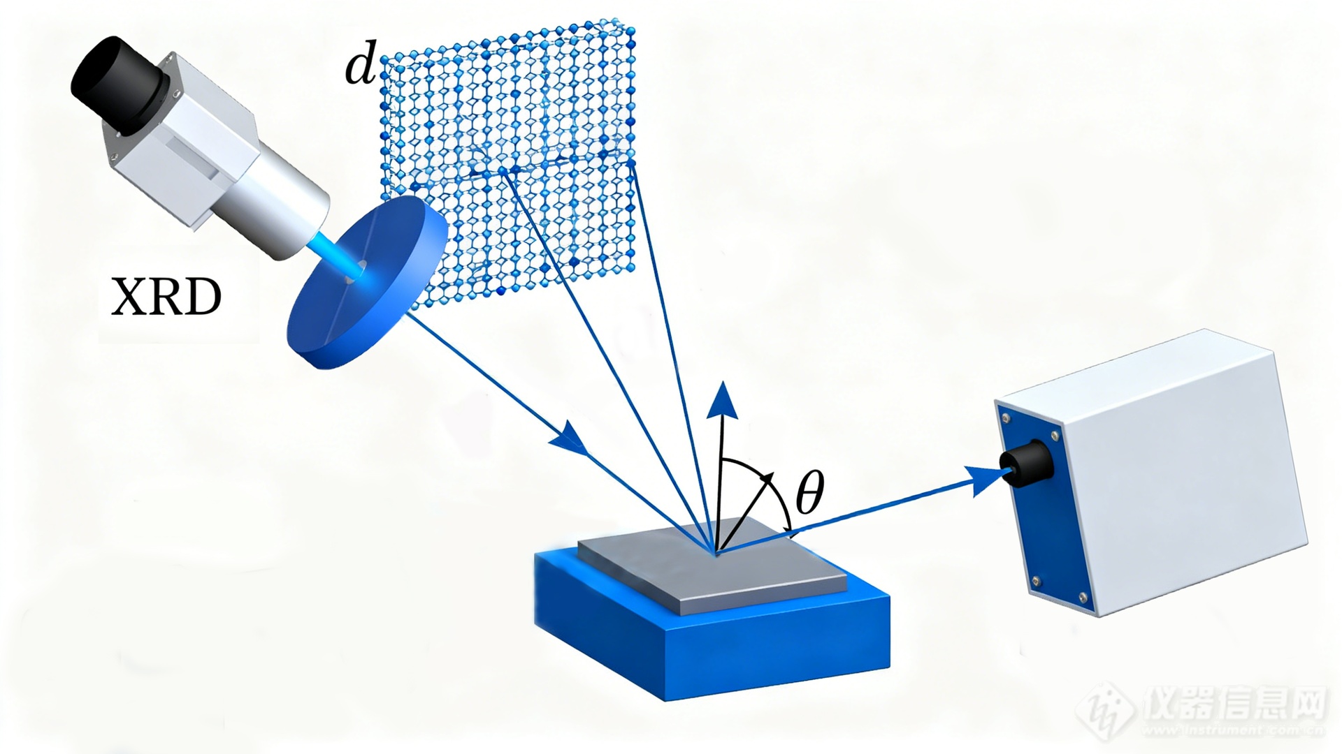
XRD schematic
The core of XRD technology is the interaction between X-rays and crystal atoms. The high-energy electron beam bombards the metal target to produce characteristic X-rays. After the sample is irradiated, the inner electrons of the atoms inside the crystal are stimulated to vibrate and generate scattered waves. Due to the periodicity of the crystal structure, scattered waves constructively interfere in specific directions, forming detectable diffraction signals. The spatial orientation and intensity of the diffraction lines directly reflect the microstructural characteristics of the crystal.
Bragg's equation is the basic theoretical basis of XRD technology: nλ=2d sinθ (where d is the crystal plane spacing, θ is the diffraction angle, n is an integer, and λ is the X-ray wavelength). This equation clarifies the necessary conditions for the generation of diffraction - only when each parameter satisfies the relationship of the equation, a strong diffraction signal will appear. However, satisfying the equation is not a sufficient condition for the generation of diffraction. The diffraction intensity is also affected by factors such as atomic distribution and crystal symmetry.
2. Technical classification
XRD technology is mainly divided into two branches: single crystal X-ray diffraction and polycrystalline X-ray diffraction.
Single crystal X-ray diffraction
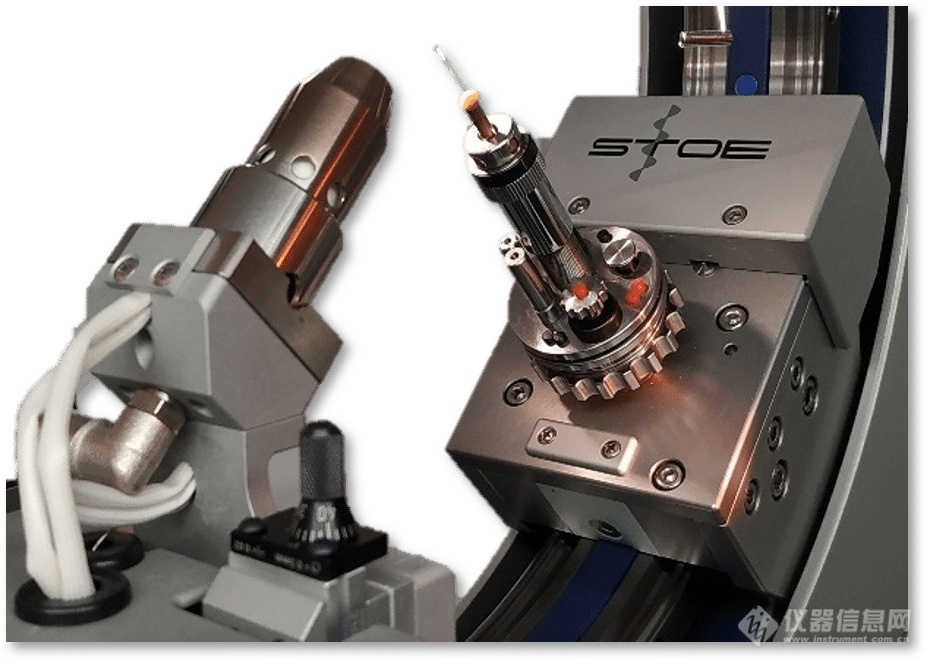
Single crystal X-ray diffractometer
The object of single crystal XRD is a perfect single crystal with a size ranging from microns to millimeters. By measuring the diffraction lattice of a crystal in three-dimensional space (reciprocal space), complete and accurate crystal structure information can be obtained, including atomic coordinates, bond lengths, bond angles, thermal vibration parameters and even electron density distribution.
Workflow: Mainly includes single crystal selection, data collection (using a four-circle diffractometer or a modern surface detector), data restoration and absorption correction, structural analysis (direct method or Patterson method) and structure refinement (least squares method).
Application and importance: SXRD is the “gold standard” for determining the absolute configuration of new compounds (such as coordination polymers, organic molecules, and natural products). In drug development, it is used to determine the precise three-dimensional structure of active pharmaceutical ingredients and their polymorphs, which is crucial for understanding drug-target interactions [2].
Polycrystalline X-ray diffraction
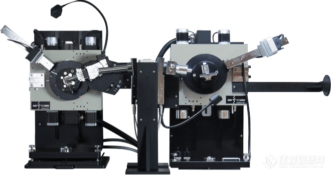
Polycrystalline X-ray diffractometer
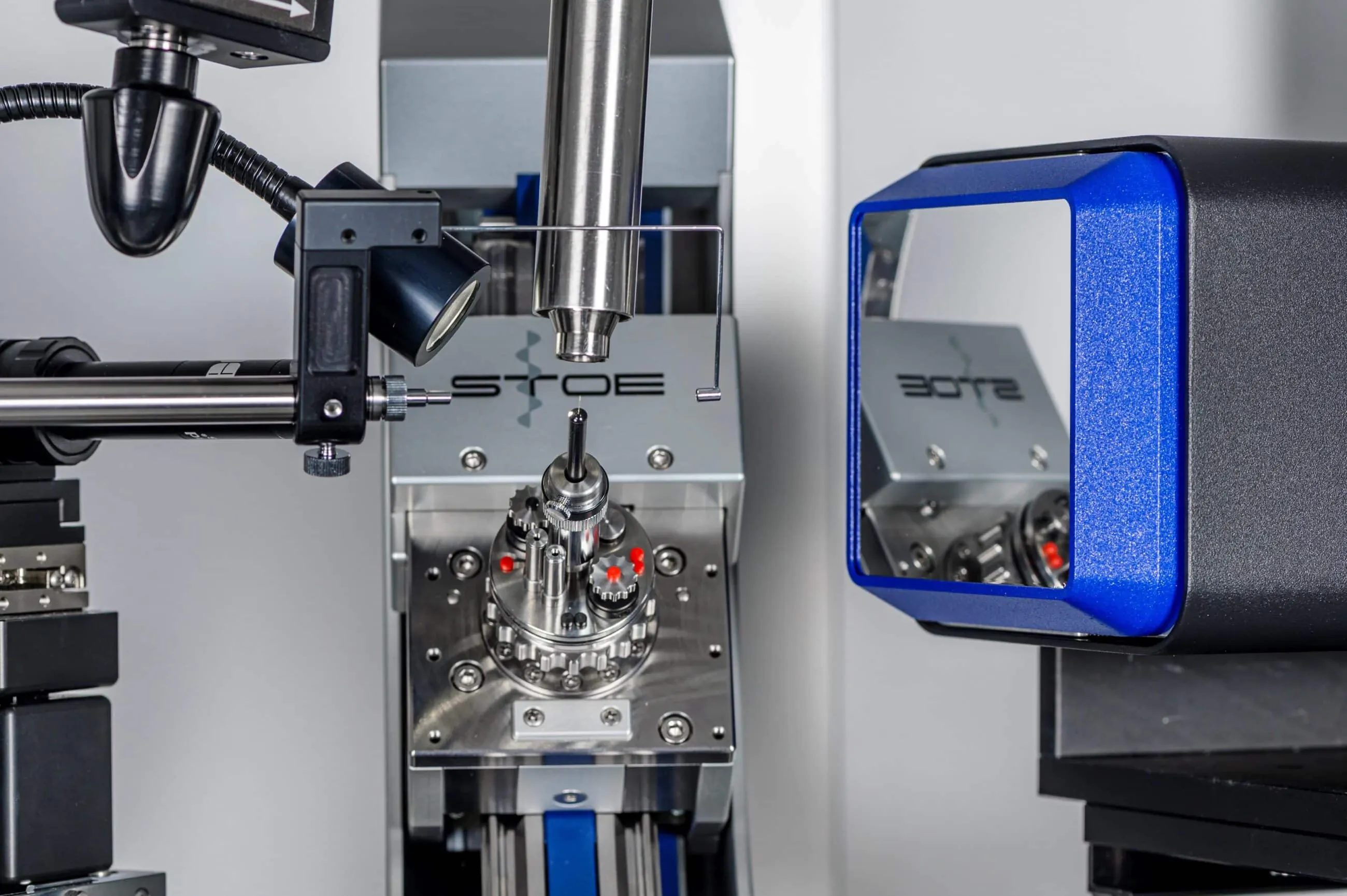
Polycrystalline X-ray diffraction (PXRD, also known as powder X-ray diffraction) uses a large number of randomly oriented tiny grains (powders) as the analysis object. As the pioneer of transmission powder XRD technology, the German STOE company has developed a transmission geometry design (such as the core technology of the STADI P series instruments) that provides a solution for the precise analysis of such samples that far exceeds the traditional reflection (Bragg-Brentano) geometry. Due to the randomness of grain orientation, three-dimensional diffraction information is usually compressed into a one-dimensional "diffraction intensity - 2θ angle" map; unlike the traditional reflection mode, the STOE transmission mode passes through X The rays penetrate evenly along the axial direction of the sample (such as loading the powder into a quartz capillary package), avoiding errors caused by sample surface flatness, particle agglomeration or tableting preparation in reflection mode, making the intensity distribution of each diffraction peak in the one-dimensional spectrum more consistent with the true orientation statistics of the grains, reducing signal distortion from the source, especially suitable for fine particles (<5μm)或易团聚样品的分析。
Qualitative and quantitative analysis of physical phases: Each crystalline substance has its unique "fingerprint" diffraction pattern. By comparing with standard databases (such as ICDD's PDF database), the phase identification of unknown samples can be achieved. In addition, based on the fact that the intensity of the diffraction peak of each phase is proportional to its content in the mixture, a variety of quantitative analysis methods can be developed.
Crystal structure refinement: Rietveld method
This is a revolutionary technology in the field of PXRD. Proposed by Hugo Rietveld in 1967, this method refines the crystal structure parameters (including unit cell parameters, atomic positions, peak shape parameters, etc.) by performing least squares fitting of the entire calculated spectrum based on the structural model and the experimental measurement spectrum [3]. Rietveld refinement makes it possible to use powder data to analyze and refine complex structures, which greatly expands the application scope of PXRD. It is especially indispensable in the research of materials where high-quality single crystals cannot be obtained (such as ceramics, catalysts, geological minerals).
Cutting edge progress and emerging technologies
With the rapid development of X-ray sources and detection technologies, XRD technology is constantly breaking through traditional limits.
1. Synchrotron radiation X-ray diffraction
The high brightness, high collimation, and adjustable wavelength X-rays provided by the synchrotron radiation light source have brought about revolutionary improvements. Its advantages include:
Ultra-high resolution and signal-to-noise ratio: can be used to study weakly diffracting samples and complex structures.
High-throughput experiments: Extremely fast acquisition speeds enable high-throughput screening of combinatorial material chips.
Small-angle X-ray scattering: used to study structural order at the nanoscale.
2. In situ X-ray diffraction
Traditional XRD provides a static "snapshot", while in-situ XRD can monitor the structural evolution of the sample in real time while it is undergoing external stimulation (such as temperature changes, pressure changes, atmosphere changes, electrochemical charge and discharge) [4]. For example, in lithium-ion battery research, in-situ XRD can reveal the phase change process of electrode materials during charge and discharge in real time, providing direct evidence for optimizing battery performance.
3. Micro-area X-ray diffraction
Combining high-brightness light sources and precision focusing optics, μ-XRD can focus the X-ray beam spot to the micrometer or even nanometer level, thereby achieving structural analysis of specific micro-regions of the sample (such as individual grains, interfaces, defects), which is extremely valuable in heterogeneous materials, failure analysis and microelectronic device characterization.
Overview of application areas
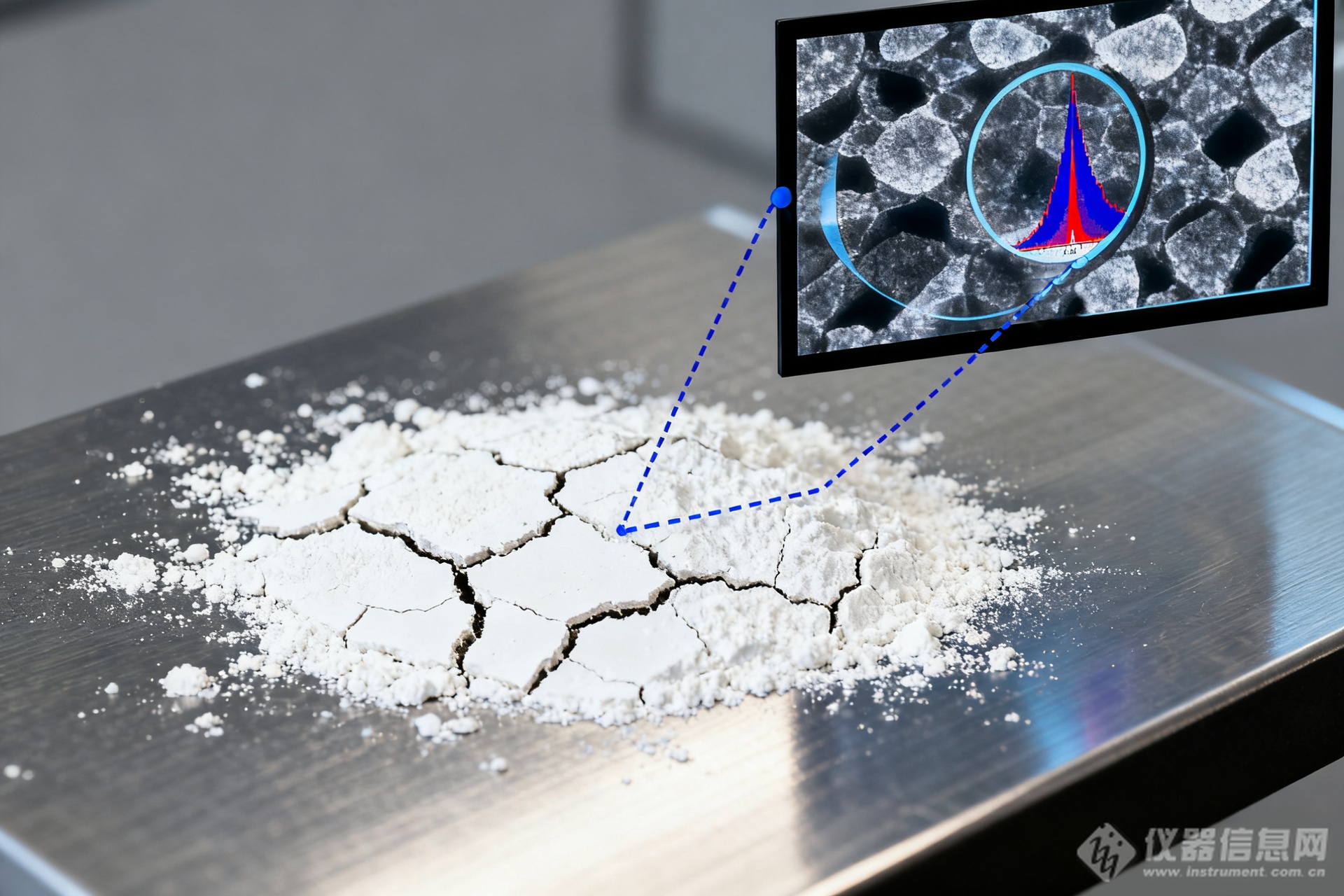
Application of XRD in the field of materials
The application of XRD technology has penetrated into many fields of science and engineering:
Materials science: discovery and characterization of new materials, phase diagram determination, residual stress analysis, texture analysis.
Pharmaceutical industry: screening and identification of drug polymorphs, quantitative analysis of active ingredients in preparations, and stability studies.
Geology: mineral identification and quantification, petrogenesis and evolutionary history research.
Archeology and cultural heritage: physical phase analysis of ancient ceramics, pigments, and metal products for dating and authenticity identification.
Life sciences: Although synchrotron radiation is more commonly used in macromolecular crystallography, laboratory XRD sources are still used for structural analysis of compounds related to protein function such as small molecule inhibitors and cofactors.
Conclusion and outlook
X-ray diffraction technology has gone through hundreds of years, and its depth and breadth are still expanding. From static structural analysis to dynamic process tracking, from macro sample statistics to micro-area single crystal analysis, XRD technology continues to provide the core structural information for scientific discovery and industrial innovation.
About STOE, a century-old German XRD manufacturing company
If the century-old development of X-ray diffraction technology is compared to a magnificent building, then those top equipment manufacturers are the most solid cornerstones and pillars of this building. Among these cornerstones, STOE, founded in 1887, is undoubtedly a monument engraved with legend. It is not only a witness to the development of the industry, but also actively shapes the direction of technological evolution with a series of groundbreaking achievements. Starting from crystal optical inspection equipment, it has been deeply involved in single crystal and powder crystal XRD since the 1960s. It not only invented the powder X-ray diffraction transmission geometry technology, but also developed the first single crystal X-ray diffraction system equipped with an open Euler bracket and a pixel detector. It is the first manufacturer to commercially apply transmission geometry to powder XRD (2 1960s); Relying on high-precision processes made in Germany, STOE's single crystal diffractometer can accurately control the dispersion spot radius below 5 microns. It is also a global authorized dealer of the International Diffraction Data Center (ICDD). Its century-old technical strength, brand reputation and global high-end customer resources have made it a recognized benchmark enterprise in the industry. The acquisition of STOE by Jingwei Gaobo (Jinwei acquired STOE, a century-old German X-ray diffractometer (XRD) manufacturing company - to comprehensively expand the single crystal and powder crystal XRD product lines), marks that Chinese capital and technology are integrating into the global high-end scientific instrument industry chain in an unprecedented way. This combination can generate more innovative and future-oriented XRD solutions, provide diversified "adsorption-thermal analysis-diffraction" solutions for new material research and production customers, and contribute new strength to the advancement of materials science in China and even the world.

References
[1] Bragg, W. L., & Bragg, W. H. (1913). The Reflection of X-rays by Crystals. Proceedings of the Royal Society of London. Series A, 88(605), 428-438.
[2] Glusker, J. P., Lewis, M., & Rossi, M. (1994). Crystal Structure Analysis for Chemists and Biologists. VCH Publishers.
[3] Rietveld, H. M. (1967). A profile refinement method for nuclear and magnetic structures. Journal of Applied Crystallography, 2(2), 65-71.
[4] Chupas, P. J., Qiu, X., Hanson, J. C., Lee, P. L., Grey, C. P., & Billinge, S. J. L. (2003). Rapid-acquisition pair distribution function (RA-PDF) analysis. Journal of Applied Crystallography, 36(6), 1342-1347.
[5] Ovchinnikov, O. S., et al. (2019). AI-enabled high-throughput X-ray diffraction analysis. MRS Bulletin, 44(9), 699-705.
View More from This Booth
Behind the rush of "third-generation Kirin battery": the "key line of defense" that delicately protects battery safety
Germany's Leibniz Institute of Catalysis uses XRD to uncover the mystery of CO₂ hydrogenation catalyst deactivation
Domestic Thermal Analysis - Empowering scientific research and industrial upgrading with sophisticated technology