

 Gold Member
 Business License
Product Details
Applications
Detailed Description
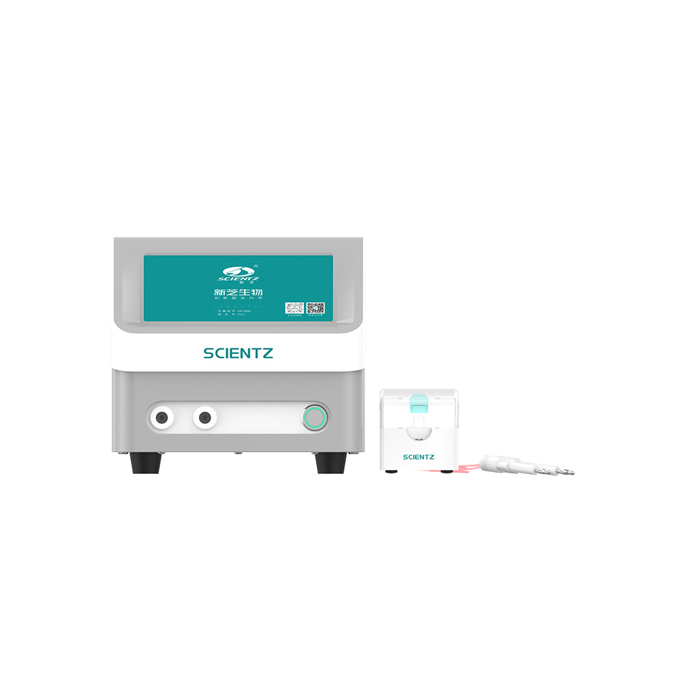
The GP-3000 Gene Transfer Instrument is composed of a main unit, gene transfer cups and dedicated connecting cables. It mainly utilizes electroporation technology to transfer DNA into competent cells, animal and plant cells, as well as yeast cells. Compared with other methods, this electroporation-based approach boasts advantages of high repeatability, high efficiency, simple operation and quantifiable control. Meanwhile, electroporation is free of genotoxicity, and has become an indispensable fundamental technique in molecular biology..
▌Working Principle
Cell electroporation (EP), also known as electropermeabilization, is an important method for introducing exogenous macromolecules (including DNA, RNA, siRNA and proteins) and small molecules into the interior of cell membranes.
Under the action of an instantaneous strong electric field, the cell membranes of cells in the solution gain a certain degree of permeability, and charged exogenous substances enter the cell membranes in a manner similar to electrophoresis. Owing to the high electrical resistance of the phospholipid bilayer of cell membranes, the voltage across the cell poles generated by the external electric current field is fully borne by the cell membranes, while the voltage distributed in the cytoplasm is negligible and there is almost no electric current inside the cytoplasm. This determines that the cytotoxicity remains extremely low during electroporation within the normal parameter range. After the electric field drives substances such as DNA into the cell membranes, the substances can only stay near the membranes, and then the intrinsic cellular mechanisms facilitate their transport to locations such as the cell nucleus.
As electroporation relies on physical means, the molecular characteristics of the cell surface exert minimal influence on the transfection effect. In contrast to chemical transfection and viral vector-mediated transfection, electroporation is applicable to all cell types and enables easy quantifiable control of the transfection process.
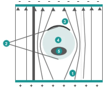
Cell electrotransfer electric field diagram
- The cell membrane is an approximate insulator, leading to current distortion in the electroporation buffer around the cell.
- In a series circuit, the higher the resistance, the greater the voltage allocated. The voltage across the cell is almost entirely borne by the cell membranes at both ends.
- Only the membrane at one cell end surface is effective for electroporation.
- The voltage distributed in the cytoplasm is extremely low. DNA immediately stops moving once it enters the cell membrane via electroporation, and subsequently diffuses slowly relying on the cell’s intrinsic mechanisms.
- The voltage across the cell nucleus is negligible, and such minimal voltage is fully borne by the nuclear membrane, with no voltage present inside the nucleus at all. This ensures electroporation is free of genotoxicity.
▌Features
High Efficiency Short transformation time, high conversion rate, small tolerance.
Intelligent Storage Experimental parameters can be stored for user convenience.
Precise Control Microprocessor-controlled pulse discharge.
Aesthetic Design Integrated design, intuitive display, simple operation.
▌Application
Food Industry | Agriculture | Veterinary Science | Bioengineering
• Electroporation of bacteria, yeast & other microorganisms
• Transfection of mammalian cells, plant tissues & protoplasts
• Cell hybridization and gene fusion
• Marker gene delivery for labeling & tracing
• Introduction of drugs, proteins, antibodies for cell structure & function research
| Specification | GP-3000 |
| Pulse Mode | Exponential decay & square wave |
| High-voltage Output Voltage | 401–3000 V |
| Low-voltage Output Voltage | 50–400 V |
| Power Supply Voltage | 100–240 VAC, 50/60 Hz |
| Continuous Discharge Times | 1–9 |
| Power | 100 W |
| Parallel-connected Resistor | 100 Ω–1650 Ω, with 1 Ω increment (50 Ω increment recommended) |
| Control Mode | Microcomputer control |
| Time Constant | RC time constant, adjustable |
| Low-voltage Capacitance | 25 μF–1575 μF, with 1 μF increment (25 μF increment recommended) |
| High-voltage Capacitance | 10–60 μF, with 1 μF increment (10 μF, 25 μF, 35 μF, 50 μF, 60 μF recommended) |
| Discharge & Interval Time | 0.1 ms–999 ms, 0.1 ms increment |
| Net Weight of Main Unit | 4.5 kg |
| Overall Dimensions of Main Unit | 30×20×20 cm |
| Packing Dimensions | 58×36×25 cm |
▌Conversion Rates for Different Strains
| Type | PulseVoltage (V) | Resistance(Ω) | Capacitance(μF) | ElectroporationCup (mm) | Type | PulseVoltage (V) | Resistance(Ω) | Capacitance(μF) | ElectroporationCup (mm) | |
| E.Coli | 1800 | 200 | 25 | 1 | S.pyogenes | 2100 | 200 | 50 | 2 | |
| E.Coli | 2500 | 200 | 25 | 2 | L.plantarum | 2000 | 400 | 25 | 2 | |
| E.Coli | 3000 | 200 | 25 | 2 | Eukaryotes | |||||
| A.tumefaciens | 2400 | 200 | 25 | 1 | S.cerevisiae | 1500 | 200 | 25 | 2 | |
| P.asruginosa | 2500 | 200 | 25 | 2 | S.pombe | 2300 | 200 | 25 | 2 | |
| S.aureus | 2900 | 100 | 25 | 2 | C.albicans | 1500 | 200 | 25 | 2 | |
| B.cereus | 1000 | 200 | 25 | 2 | P.pastoris | 2000 | 200 | 25 | 2 | |
Note: Due to variations in experimental conditions across different labs,the above parameters are for reference only.
Example Experiments
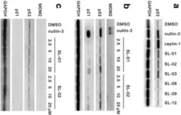
1、LIJ, ZHANG S, GAO L, et al. A cell-based high-throughput assay for the screening of small-molecule inhibitors of p53-MDM2 interaction [J]. Journal of biomolecular screening, 2011.
2、ZHANG SY, LIJ, XIE X. Discovery and characterization of novel small molecule agonists of G protein-coupled receptor 119 [J]. Acta pharmacologica Sinica, 2014.
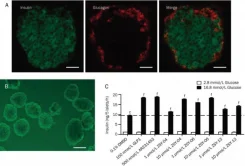
| [1]LIJ and colleagues analyzed the small molecule inhibitors of p53-MDM2 protein interactions based on high-through- put screening. | 2、ZHANG SY and colleagues discovered and identified a unique G-protein-coupled receptor 119 agonist. [2] |
 |  |
Product Overview
The GP-3000 Gene Transfer Instrument consists of a main unit, gene transfer cups and special connecting cables. It mainly adopts the electroporation technology to transfer DNA into competent cells, animal and plant cells as well as yeast cells. Compared with other methods, the method using this instrument features prominent advantages including high repeatability, high efficiency, easy operation and quantifiable control. It supports two pulse modes: exponential decay and square wave. The instrument is equipped with a high-voltage capacitor ranging from 10 to 60 μF with a step of 1 μF, a low-voltage capacitor ranging from 25 μF to 1575 μF with a step of 1 μF, and a parallel-connected resistor ranging from 50 Ω to 1650 Ω with a step of 1 Ω.
Send an inquiry
Recommended Products
