
 Gold Member
 Business License
qPCR is always "late", and dPCR also overturns? Maybe the PCR inhibitor is causing trouble
Everything is normal with the standard product. Change to soil, feces,whole blood, plant and food extracts, Cq is suddenly delayed;
The amplification curve is good but the fluorescence is weak, but there is a target band in the agarose gel;
The overall qPCR quantification is low, and the standard curve is unstable;
Switching to dPCR, I thought that "digital typing is not afraid of suppression", the results sectiondroplets/The micropores are not bright and the absolute copy number is also low.
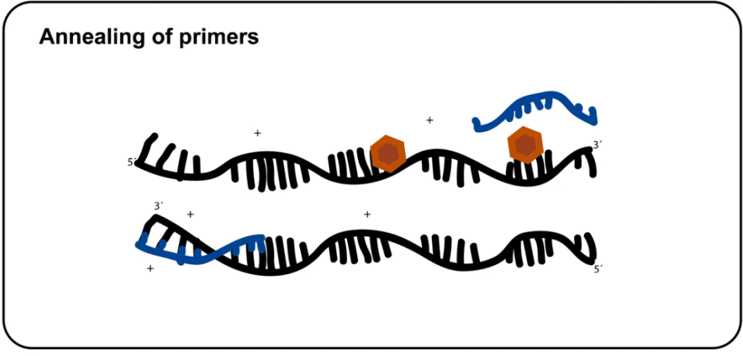
Behind these problems, a large part isPCR inhibitors at work. Analytical and Bioanalytical Chemistry This review combines qPCR, dPCR,MPSSystematic review of the inhibitory mechanism: PCR is not just as simple as “adding Template + Master Mix”, it also relies on in vitro DNA polymerization,Primer annealing, denaturation and renaturation, and fluorescence detection; as long as certain molecules in the matrix hit any of these links, the results will be distorted.
In the review, the key sub-reactions of PCR are drawn as a chain: template denaturation → primer binding to single strand → polymerase extension → product accumulation → fluorescence reading. so-calledPCR inhibitors, generally refers to any substance from the sample matrix, target cells, and extraction reagents that can interfere with in vitro polymerization or fluorescence signals.
There are three common types of action points:
1. polymerase itself
Reduce the activity of Taq/thermostable DNA polymerase and reduce the overall amplification efficiency. When the early cycle is affected, Cq is pushed back; when the entire cycle is affected, amplification may not occur at all. Humic acid, hemoglobin, methemoglobin, bile salts, phytic acid, phenols, etc. may affect the polymerase.
2. Nucleic acid/primer annealing
Binding to ssDNA or dsDNA blocks primer access to the template, especially in the first few cycles. IgG is typical: it binds to genomic ssDNA, interferes with primer annealing, and manifests as Cq delay.
3. Fluorescence detection
It does not necessarily cause amplification failure, but it can make the instrument "see clearly". Humic acid and hemoglobin can form static quenching complexes with free dyes, suppressing the signals of SYBR Green I, EvaGreen, ResoLight, ROX, etc.; DTT can quench passive reference dyes in some systems. This type is very hidden: amplification has actually occurred, and the bands can be seen in gel electrophoresis, but the qPCR curve peaks late and has a low platform; in dPCR, the passive reference is suppressed, and the yin and yang thresholds are set biased.
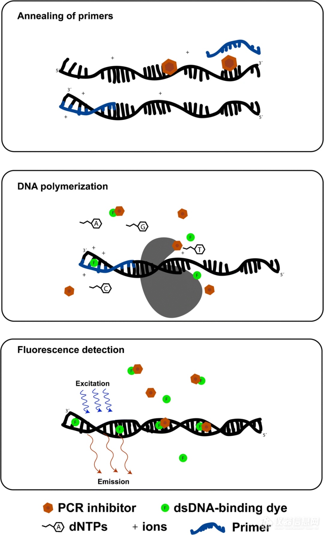
Figure 1. Schematic diagram of the key reaction steps of PCR. PCR inhibitors may affect any of these reactions, for example by interfering with primer annealing, affecting DNA polymerase activity, or impairing fluorescence detection.
qPCR relies on Cq value (quantification cycle number) and standard curve for absolute or relative quantification, and the interference of inhibitors on early amplification kinetics is fatal.
1. Efficiency distortion:In the presence of inhibitors such as soil humic acid and fecal bile salts, the slope of the amplification curve changes, and the calculated efficiency may directly fall out of the acceptable range of 90%-110%, resulting in quantitative results that are too high or too low.
2. Fluorescence artifact:If you find that the amplification curve cannot rise but there is product when running the gel, it is most likely that the fluorescence has been quenched. Especially when using dyes such as SYBR Green, humic acid not only inhibits enzymes, but also severely reduces fluorescence intensity.
3. Internal standard rescue:Adding an internal standard (IAC) to the qPCR system is an effective way to identify inhibition. If both the internal standard and the target are delayed at the same time, it means that the inhibitor is causing trouble, not the target sequence itself.
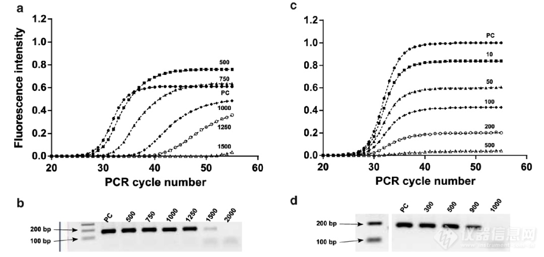
Figure 2. Example of PCR inhibition in qPCR. a. As the amount of fulvic acid that inhibits amplification increases, the amplification curve obtained is analyzed. b. Agarose gel electrophoresis results corresponding to the amplification reaction in Figure a. c. As the amount of humic acid added to quench fluorescence increases, analyze the amplification curve obtained. d. Corresponds to the agarose gel electrophoresis results of the amplification reaction in Figure c. The numbers in the figure represent the amount of humic acid or fulvic acid added to the reaction system. PC (positive control) represents the reaction in which water replaced the PCR inhibitor. Detection uses EvaGreen dye.
dPCR: More resistant to infection, but not a "gold medal for immunity from death"
dPCR divides the reaction system into tens of thousands of microdroplets/microchambers to perform endpoint interpretation without relying on Cq values. Therefore, it does tolerate mild inhibition better than qPCR. But the review clearly points out: dPCR is not a panacea.
1. Partition dilution effect:Although partitioning reduces the inhibitor concentration, if the inhibition is too strong, some droplets will still fail to amplify, resulting in a decreased positivity rate and an underestimate of the absolute copy number.
2. Enzymes vary greatly:The tolerance of different polymerases to inhibitors varies widely. The article mentioned that by using specific tolerance enzymes or buffers, dPCR's tolerance to humic acid can be increased dozens of times.
3. The reference fluorescence is suppressed:dPCR interpretation of Yin and Yang requires setting a fluorescence threshold. If hemoglobin or humic acid in the sample quenches passive reference dyes such as ROX, it will cause errors in threshold normalization and misjudge weak positives as negatives.
Environmental Soil/Water:Humic acid, fulvic acid. A two-pronged approach, it not only inhibits enzymes but also quenches SYBR/EvaGreen fluorescence.
Blood/Plasma:Hemoglobin, methemoglobin, IgG. Hemoglobin directly poisons the polymerase and quenches the dye; IgG sticks to the DNA like "glue" and prevents primer binding.
Feces/intestine:Bile salts, phytic acid, polysaccharides. Mainly inhibits polymerase activity, resulting in low amplification efficiency.
Plants/Food:Polyphenols, tannins, polysaccharides, pigments. Polyphenols bind to proteins after oxidation, and polysaccharides increase viscosity and co-precipitate DNA.
In the face of inhibitors, the traditional "infinite dilution" strategy often discards precious low-abundance templates. The following combinations of punches are recommended:
1. Enzyme replacement is the first choice:Don’t just focus on “high fidelity”, look for polymerases that are “inhibition resistant”. Select blood-resistant enzymes for blood samples and humate-resistant enzymes for soil.
2. Use additives wisely:
BSA (bovine serum albumin): can effectively adsorb some inhibitors and rescue the inhibited polymerase, but is ineffective in dye quenching.
T4 gp32/Dehumicants: For environmental samples, try using specialized inhibitor removers.
3. Adjust detectionmethod:If SYBR Green is always quenched, try switching to a hydrolysis probe (such as TaqMan), which usually suffers less interference from matrix fluorescence.
4. Shorten the amplicon:In dPCR and suppressed qPCR, try to control the amplicon length to 70-120bp. Short fragments are more likely to be successfully amplified in adverse conditions than long fragments.
References:
Sidstedt M, Rådström P, Hedman J. PCR inhibition in qPCR, dPCR and MPS—mechanisms and solutions. Analytical and Bioanalytical Chemistry, 2020, 412:2009–2023.
Related products

Swipe left or right to see more →
View More from This Booth
Jie Laimei's three-dimensional digital system was unveiled at the National Chinese Medicinal Material Germplasm Resource Bank, and Hunan Satellite TV reported it!
qPCR is always "late", and dPCR also overturns? Maybe the PCR inhibitor is causing trouble

Good News | Jie Laimei Technology was awarded the 2026 Chengdu Enterprise Technology Center
Application Case | Jie Laimei's three-dimensional digital specimen rapid construction system empowers ginseng morphological identification and digital specimen library construction
Jie Laimei digital PCR: bringing droplet matrix dPCR solution to complex matrix detection of wastewater
Recommended Products