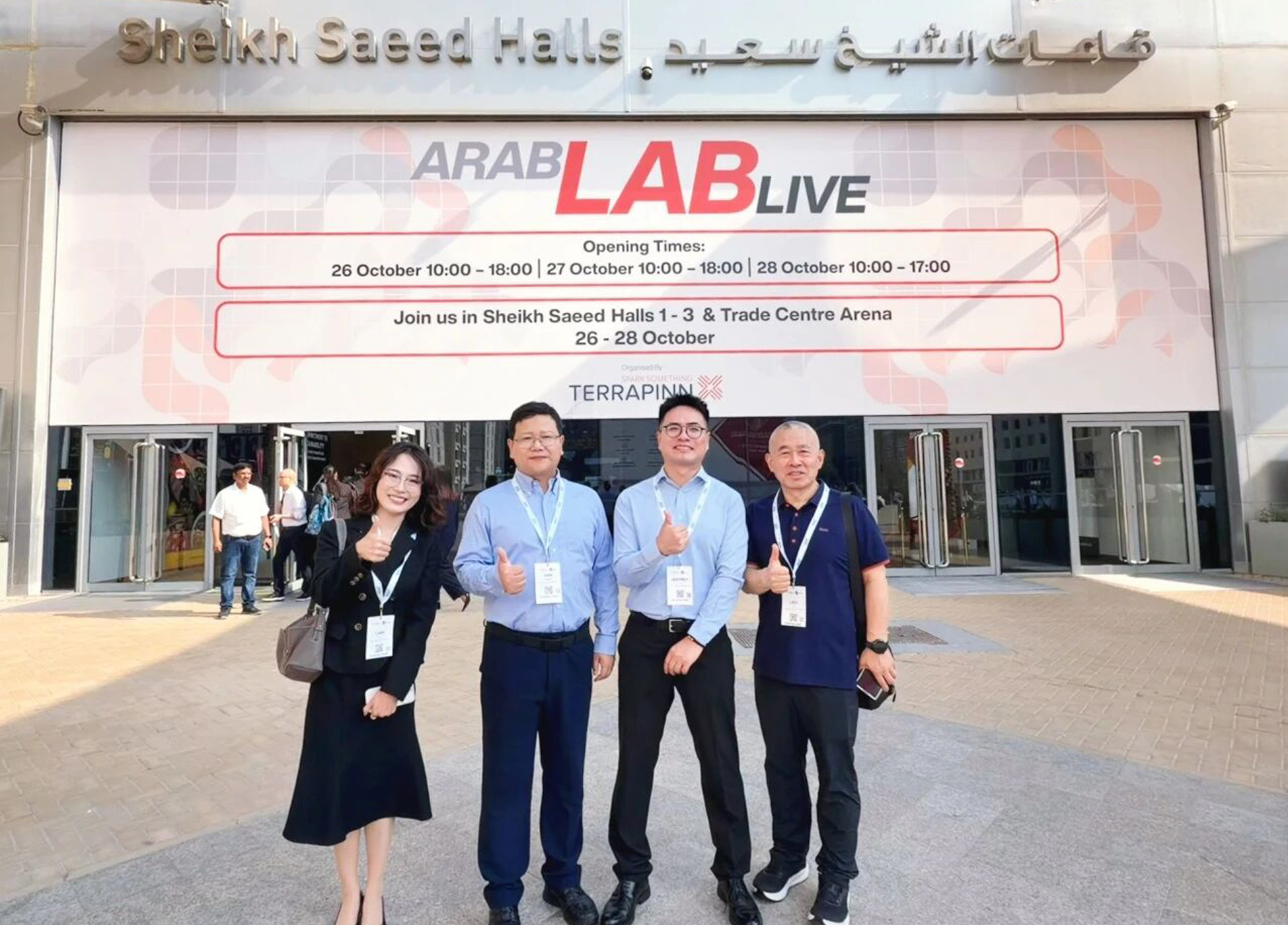
Being close to users continues from every meeting, Haineng goes to ARABLAB again
Company News
Oct 30, 2026

EBIT Life Sciences Content Team
2026-09-07
Western In the last step of Blot (WB), many people will encounter the same choice: use fluorescence method or chemiluminescence method?
Let me start with the conclusion: no one method is better in all scenarios. If your core requirement is to detect low-abundance proteins and use the mature HRP antibody system, chemiluminescence is usually more suitable; if you need to distinguish multiple targets simultaneously on the same membrane, or want to obtain a wider and more stable quantitative window, fluorescence methods are usually more advantageous. What really affects the choice is the purpose of the experiment, protein abundance, quantitative requirements, existing antibodies and instrument conditions.
Fluorescence and chemiluminescence methods, where do the signals come from?
Chemiluminescence methods usually use HRP-labeled secondary antibodies. HRP catalyzes the luminescent substrate to produce a light signal, which is then recorded by film or digital imaging equipment. It involves an enzymatic amplification process, making it particularly suitable for capturing low-abundance targets; however, the signal will vary with substrate reactions, and acquisition timing and exposure conditions will affect the results.
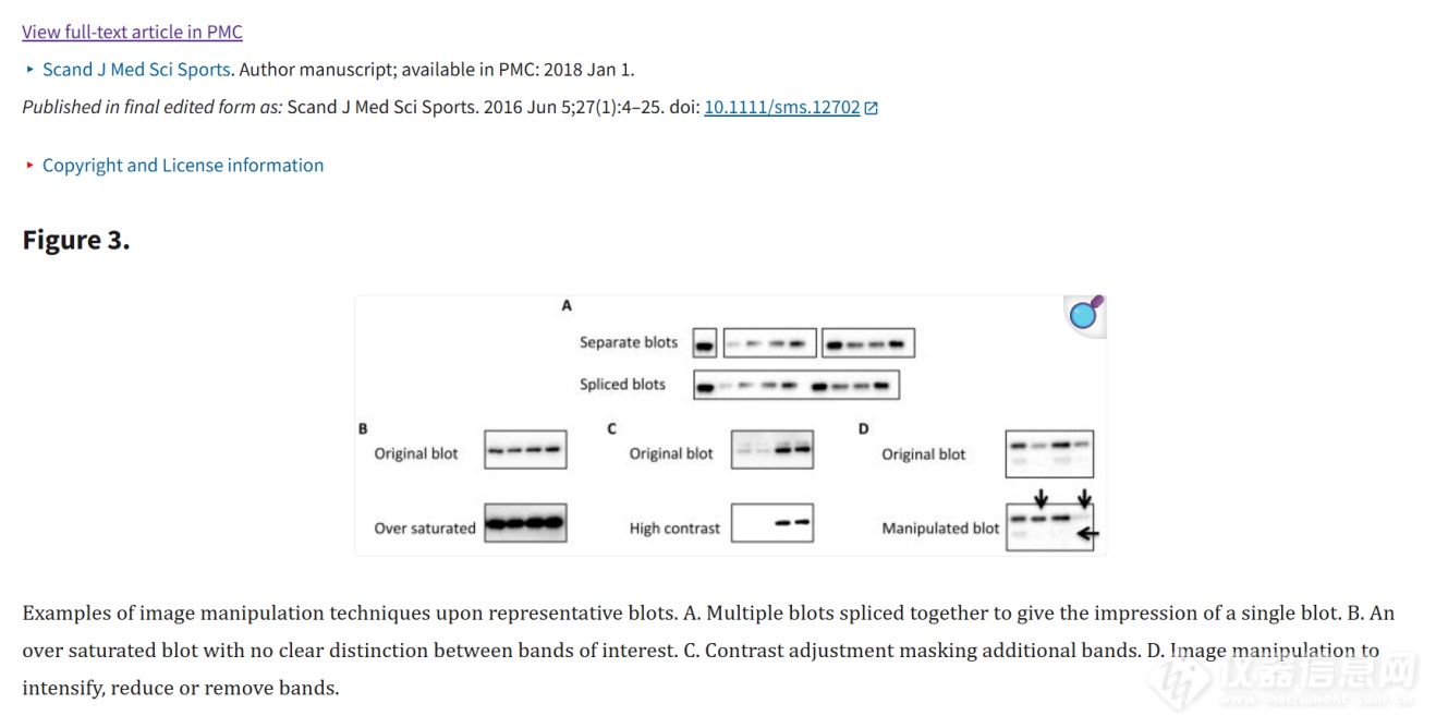
The band intensity is suitable for relative quantification only when the WB signal is within a verified linear interval; a signal that is too weak may be overwhelmed by the background, and a signal that is too strong may lose discrimination due to saturation. For principles, please refer to the WB technology review by Bass et al.
The fluorescence method uses secondary antibodies with fluorescent groups. The imaging system excites the fluorophore at a specific wavelength and collects its emitted light. Different spectral channels can record different targets respectively, so they are more suitable for multiplex detection; their signals are usually more stable than chemiluminescence, but light protection, channel crosstalk and background fluorescence need to be controlled.
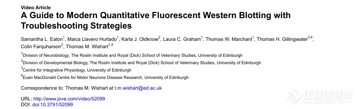
Multi-channel fluorescent WB can distinguish different targets on the same membrane, reducing differences caused by membrane separation or stripping counterstaining; however, dye spectrum, antibody host, channel crosstalk and linear range still need to be verified in advance. For principles, please refer to Eaton et al.
Six key dimensions, understand two methods at once
Contrast Dimensions | Chemiluminescence | Fluorescence method | |
signaling mechanism | HRP and other enzymes catalyze the substrate to emit light, and there is signal amplification | Fluorescent groups emit light of a specific wavelength when excited | |
Sensitivity | Often more suitable for low-abundance or very low-abundance targets | Good sensitivity, actual performance depends on dye, equipment and background | |
multiplex testing | Conventional HRP single channel mainly focuses on a single target; multiple targets often need to be separated or stripped and then counterstained. | Different spectral channels can be used for multiplex detection of the same membrane. The number of channels depends on the instrument and dye combination. | |
Linear vs. quantitative | Enzyme reactions, substrate consumption, and saturation narrow the quantifiable interval, but gradient validation still allows for reliable relative quantification. | Typically has a wider linear dynamic range, making it easier to perform same-membrane normalization and multi-target relative quantification. | |
Signal stability | The signal changes with the reaction time and needs to be collected in time within the appropriate window. | Can usually be scanned again when protected from light and stored properly, but photobleaching may still occur | |
Costs and Thresholds | The HRP antibody system is mature and popular, and the cost of routine use is usually low. | Matching excitation light sources, filters and fluorescent secondary antibodies are required, and multiple experiments also need to control cross-reaction and cross-color. |
What needs to be emphasized is that"Wider dynamic range" does not mean automatically obtaining accurate data. Regardless of the method used, confirm that the target band is in the unsaturated linear region of the detector and verify the loading amount, antibody concentration, and exposure parameters. Quantitative WB also requires appropriate normalization methods, sufficient biological replicates, and transparent image processing records.
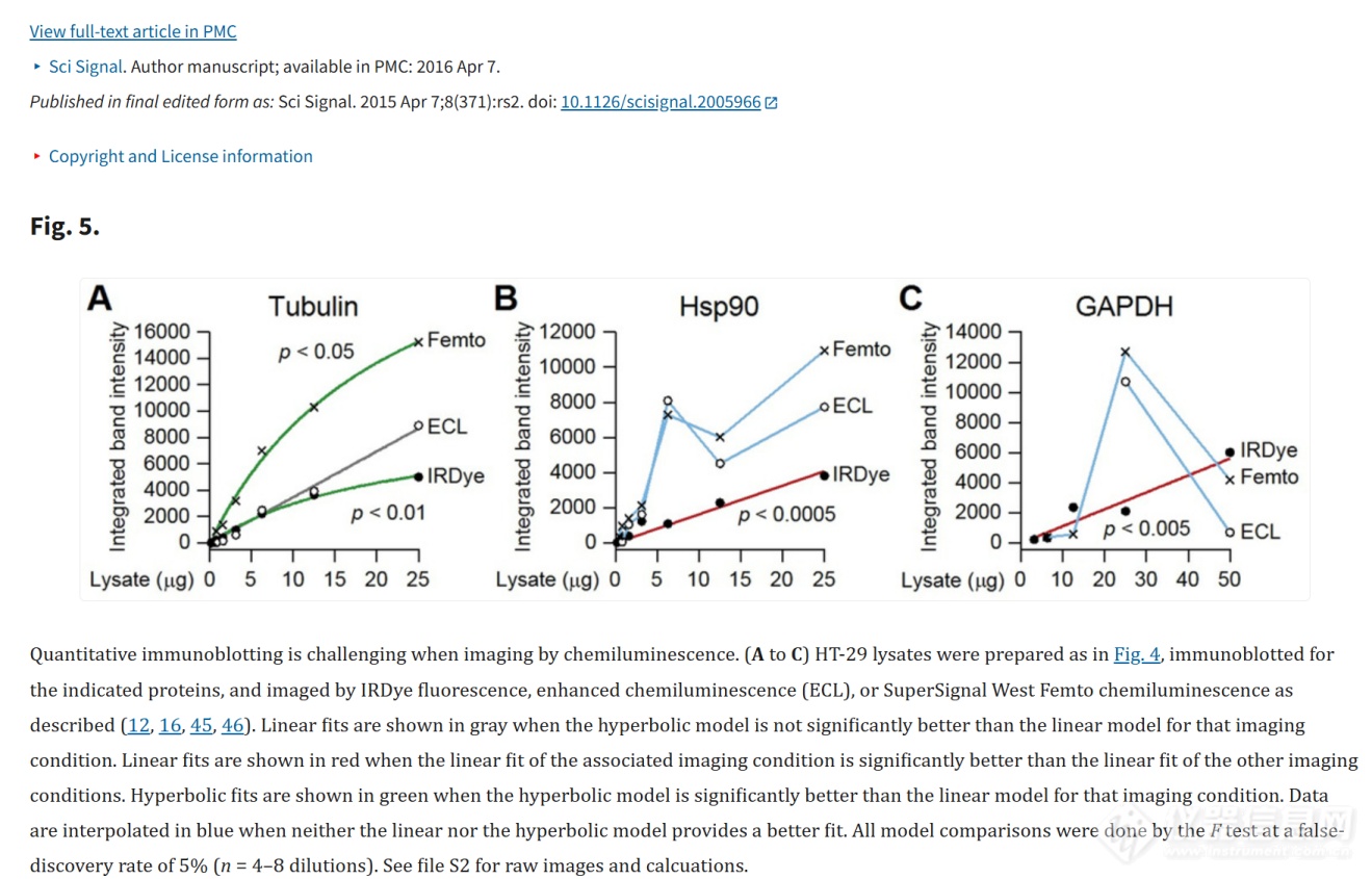
The interpretable relationship between the detection signal and the true protein amount can only be maintained within a validated linear interval. Nonlinear amplification may overestimate changes, and detection saturation may underestimate changes. Schematic diagram based on Janes’ quantitative immunoblotting studies
Under what circumstances is chemiluminescence preferred?
The following scenarios are generally more appropriate to start with chemiluminescence:
• Target protein abundance is low, such as partially phosphorylated proteins, targets after co-immunoprecipitation, or weak signals in precious samples;
• It mainly detects one target protein, and there is no need for multiple imaging of the same membrane;
• The laboratory has established a stable HRP antibody and ECL substrate system, hoping to reduce method migration costs;
• The budget is limited, and the existing workflow is mainly based on conventional WB qualitative or relative quantitative.
The main risk of chemiluminescence is not “not quantifiable”;More susceptible to losing quantitative value due to exposure or signal saturation. If the strong band has reached the upper limit of detection, no matter how bright it is, it will not produce a distinguishable grayscale difference; if the weak band falls into the background noise, "not captured" does not mean "no expression". Therefore, original images should be retained and linear intervals confirmed through short exposures, gradient loading, or series of exposures; uncropped, minimally processed original images should also be saved and provided for publication.
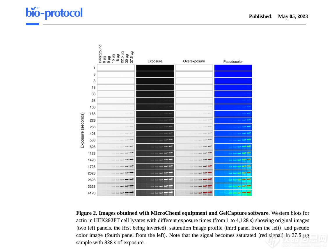
Chemiluminescent WB allows for relative quantification, but saturation should be identified through a series of exposures and a common linear interval selected for all samples to be compared. The principle refers to the chemiluminescence quantitative process proposed by Cañada-García and Arévalo.
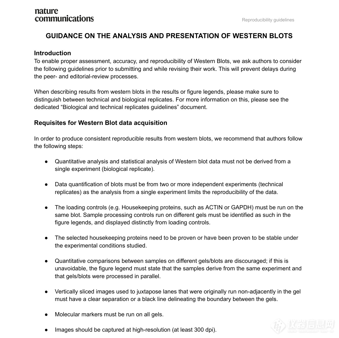
Regardless of whether you choose fluorescence or chemiluminescence, reliable WB data requires independent repeats, appropriate controls, unsaturated acquisition, complete raw images, and transparent processing records. Requires WB data and image specifications compiled from Nature Portfolio
Under what circumstances is fluorescence preferred?
The following requirements can better utilize the advantages of fluorescence method:
• Want to detect target protein, internal control, or total protein and phosphorylated protein simultaneously on the same membrane;
• Multiple targets have similar molecular weights and are difficult to distinguish in the same channel using conventional chemiluminescence;
• It is necessary to reduce the signal loss and inter-membrane differences caused by stripping counterstaining;
• The research focuses on the relative changes between groups, hoping to obtain a wider linear detection window;
• Very limited sample, would love to get more information from the same film.
The price of the fluorescence method is that the experimental design is more demanding. When performing multiplex detection, it is best for the primary antibodies to come from different hosts; the fluorescent secondary antibodies and excitation/emission channels need to be correctly matched; and attention should also be paid to the autofluorescence of membranes, blocking solutions, and contaminants. When target abundances differ significantly, one should also assign brighter dyes to weaker targets and verify that there is no significant crosstalk between channels.
A decision table: choose according to the purpose of the experiment
your primary goal | Prioritize | Reason for selection | |
Detect low-abundance single targets as much as possible | chemiluminescence | Enzymatic signal amplification, often with higher ultimate sensitivity | |
Detect 2 or more targets on the same membrane | fluorescence | Different spectral channels can simultaneously distinguish multiple signals | |
Make a more stable relative quantitative | Fluorescence priority while verifying linear region | Typically wider linear range, but still requires standard curve and unsaturated acquisition | |
Follow mature HRP processes and control costs | chemiluminescence | The antibody and substrate systems are mature and the switching cost is low | |
Samples are precious | Depends on goal | For multi-target information, fluorescence is selected; for extremely low abundance detection, chemiluminescence can be prioritized | |
There are both low-abundance targets and multiple quantification is required. | Do a preliminary experiment first | Compare signal-to-noise ratio, linear range and repeatability before deciding on a solution |
Why is imaging equipment still critical after choosing chemiluminescence?
The high sensitivity of chemiluminescence is only an advantage on the "signal generation end". Whether the light emitted by the film can be fully collected and whether strong and weak bands can be distinguished under the same collection conditions also depends on the optical path, detector and collection method of the imaging system.
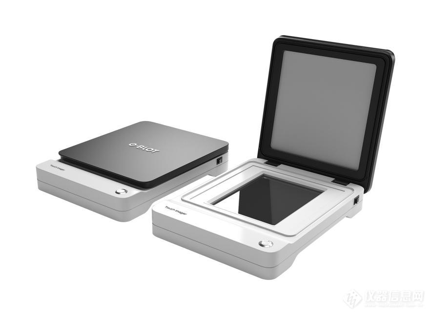
Touch Imager electronic tablet imager
Touch Imager is a contact imaging device for chemiluminescent samples such as WB. Different from the imaging path that collects signals from a long distance through the lens, it allows the film to be closely attached to the large-size photosensitive chip, reducing the loss of light in the propagation and lens links. For users, the value of this design is not an abstract parameter, but increases the chance of weak signals being recorded and reduces the burden of repeated test shots to find suitable exposure conditions.
In actual workflow, Touch Imager provides automatic, manual single and manual multiple imaging methods; supports collecting pre-stained markers and merging them with chemiluminescence images; supporting Touch Viewer can perform strip grayscale analysis, background subtraction and result export. Its wide signal acquisition range helps to present strong and weak bands simultaneously, butAny quantitative conclusions should still be premised on unsaturated, linearly verified data。
If you decide to use chemiluminescence, especially if you often face the problems of weak signal, coexistence of strong and weak bands, or time-consuming exposure, Touch Imager provides a solution for the chemiluminescence collection process. The safest way to select is to bring your own film and compare the signal-to-noise ratio, saturation, exposure time and repeatability under the same sample and lighting conditions.
Frequently Asked Questions (FAQ)
1. Can chemiluminescence WB only do semi-quantitative testing?
no. Chemiluminescence allows reliable relative quantitation, but only if the signal of interest is within a validated linear interval, the image is not saturated, and appropriate normalization and replicate designs are used. Direct quantification based on only one exposure picture that “looks suitable” is risky.
2. Is it necessary to choose fluorescence when doing quantitative WB?
uncertain. Fluorescence generally provides a wider, more stable quantification window, but chemiluminescence can also support relative quantification after specification optimization. The selection should be based on target abundance, desired detection range, antibody resources, and equipment conditions.
3. Can fluorescence methods be used to detect multiple targets simultaneously?
Conditions need to be met: the instrument has corresponding channels, the dye spectrum is separable, the combination of primary antibody host and secondary antibody is reasonable, and cross-reaction and cross-color have been eliminated. Multiplexing capabilities cannot be automatically obtained by simply changing a fluorescent secondary antibody.
References
1. Bass JJ, et al. An overview of technical considerations for Western blotting applications to physiological research. Scandinavian Journal of Medicine & Science in Sports. 2017.(Reviewed by the University of Nottingham research team)
2. Eaton SL, et al. A Guide to Modern Quantitative Fluorescent Western Blotting with Troubleshooting Strategies. Journal of Visualized Experiments. 2014.(Methodology paper by the University of Edinburgh research team)
3. Janes KA. An Analysis of Critical Factors for Quantitative Immunoblotting. Science Signaling. 2015.(Methodology paper by the University of Virginia research team)
4. Canada-Garcia D, Arevalo JC. A Simple, Reproducible Procedure for Chemiluminescent Western Blot Quantification. Bio-protocol. 2023.
5. Nature Portfolio. Western Blot guidelines.(WBOriginal image, cropping and data presentation requirements)
6. Yibit Life Sciences (Shanghai) Co., Ltd.. Touch ImagerProduct introduction.
7. Yibit Life Sciences (Shanghai) Co., Ltd.. 《TOUCH IMAGEROperating InstructionsVersion 2.4,2026-04-21。
Content boundary description: This article is used as a reference for scientific research method selection and equipment selection, and does not replace specific antibodies, reagents, and instrument instructions. The performance of different samples, antibodies, membranes, substrates and equipment may be different. It is recommended to complete gradient and repeatability verification before formal experiments.




When you visit any website, it may store or retrieve information on your browser, mostly in the form of cookies. This information might be about you, your preferences or your device and is mostly used to make the site work as you expect it to. The information does not usually directly identify you, but it can give you a more personalized web experience. Because we respect your right to privacy, you can choose not to allow some types of cookies. Click on the different category headings to find out more and change our default settings. However, blocking some types of cookies may impact your experience of the site and the services we are able to offer.