

 Gold Member
 Business License
Customer Literature | Research on surface structure analysis and osseointegration performance of titanium alloy based on nano-cerium oxide coating
Recently, the research team of Tangdu Hospital Affiliated to Xi'an Medical University published an article in an internationally renowned journal.《BioactiveMaterials》An important research result was published. This study combines in vivo and in vitro experiments with a micro-focus CT imaging system to reveal the application of "nanozyme-like" cerium oxide coatings with multiple free radical scavenging activities in bone immunomodulation and vascularized osseointegration, especially for the regulation of the immune microenvironment at the bone-implant interface. The first author of this study isTeacher ShusenBao。
Research summary
In response to the problem of postoperative aseptic loosening in the treatment of orthopedic diseases, osseointegration is an overall process that occurs at the bone-implant interface, covering immune regulation (such as macrophage polarization), angiogenesis, and osteogenesis in sequence. In order to achieve early, rapid and ideal osseointegration, this study synthesized cerium dioxide (CeO₂₋ₓ) coatings of different nanoforms (nanocones, nanopolyhedra and nanoflowers, abbreviated as NC, NP and NF, respectively) on the surface of titanium alloy (TC4) through hydrothermal method. These coatings have "nanozyme-like" activity and can scavenge a variety of free radicals and regulate bone immunity. In vitro cell experiments show that nano-CeO₂₋ₓ-coated TC4 can not only induce the polarization of RAW264.7 cells toward the M2 phenotype, but also promote angiogenesis and vascularization of endothelial cells, while promoting the differentiation and mineralization of osteoblast precursor cells. In a rat femoral condyle model, improvements in M2 polarized macrophages, angiogenesis, and bone regeneration were further demonstrated. Among the three nanoforms, NF showed the best osseointegration effect. RNA sequencing and mechanism exploration revealed that inhibiting the PI3K-AKT signaling pathway is critical for the immune regulatory ability of NF. This study provides promising ideas for the development of immune modulation in orthopedic implants.
Materials collection and research methods
Nanocoating preparation and characterization: Titanium alloy (TC4) discs and screws were sandblasted and acid-etched, and then hydrothermally treated at 120°C, 180°C, and 210°C respectively to prepare cerium oxide (CeO) with nanocone (NC), nanopolyhedron (NP), and nanoflower (NF) morphology.₂₋ₓ)coating. The morphology, structure, chemical composition and hydrophilicity of the coating were characterized through techniques such as scanning electron microscopy (SEM), transmission electron microscopy (TEM), X-ray photoelectron spectroscopy (XPS) and contact angle measurement.
Cell experiment sampling: RAW264.7 cells, HUVECs and MC3T3-E1 cells were seeded on bare titanium and nano-CeO respectively.₂₋ₓCoated TC4 disc. After 3 days of culture, the cells were collected for immunofluorescence staining, enzyme-linked immunosorbent assay (ELISA), cell migration experiment, and osteogenic differentiation experiment to evaluate the effect of the nanocoating on cell behavior.
Sampling for in vivo experiments: Nano-CeO implanted in the femoral condyle of SD rats₂₋ₓAfter coating the screws, femoral condyle tissue samples containing the screws were taken out at 1 week, 2 weeks, 4 weeks and 6 weeks respectively forMicro-CTanalyze, VanGieson (VG) staining, Masson staining and immunofluorescence staining to evaluate osseointegration, angiogenesis and inflammatory response.
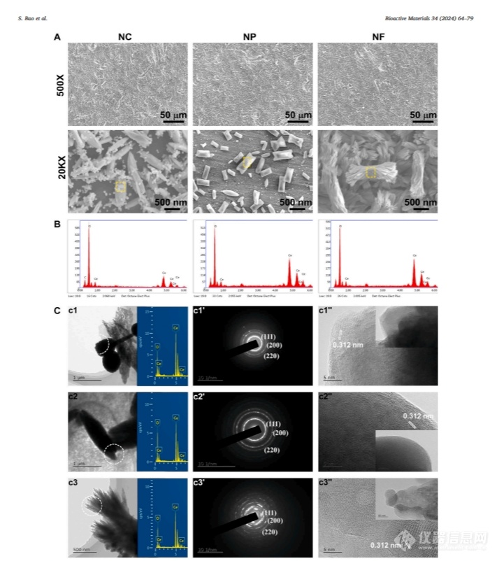
Figure 1. Structural characterization of cerium oxide coated macro/micro rough titanium alloy (TC4) in NC, NP and NF shapes. (A) Low- and high-magnification scanning electron microscopy (SEM) images. (B) Energy dispersive spectroscopy (EDS) spectrum of the yellow marked region in (A). (C) Transmission electron microscopy (TEM) images of NC, NP, and NF shaped cerium oxide scraped from the corresponding coatings, including brightfield images, inserted energy dispersive (EDX) spectra, selected area electron diffraction (SAED) patterns, and high-resolution transmission electron microscopy (HRTEM) images.
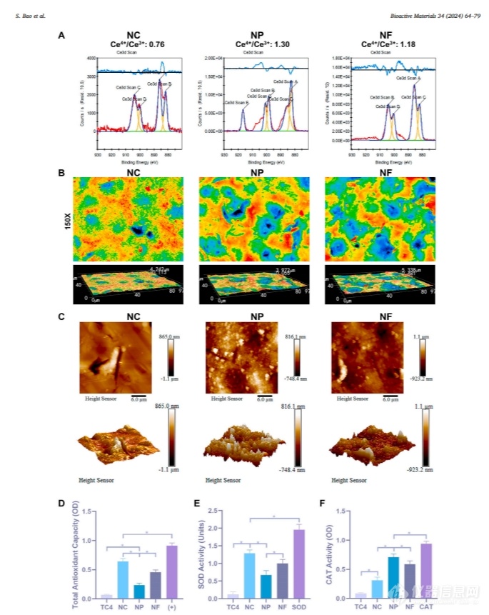
Figure 2. (A) X-ray photoelectron spectroscopy (XPS) peak differentiation of cerium (Ce) 3d; (B) Laser scanning confocal microscopy (LSCM) image; (C) Atomic force microscopy (AFM) image; (D) Total antioxidant capacity; (E) Superoxide dismutase (SOD) activity; (F) Catalase (CAT) activity. The test objects are NC, NP and NF shaped cerium oxide coatings (+: positive control). *p<0.05。
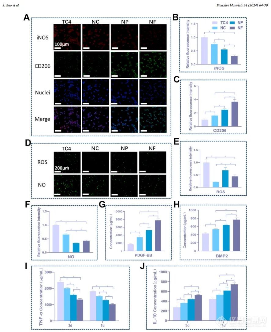
Figure 3. Phenotypic polarization and free radical scavenging ability of RAW264.7 cells on TC4, NC, NP and NF: (A) Fluorescence images of inducible nitric oxide synthase (iNOS, red), differentiation cluster 206 (CD206, green) and nucleus (blue marker) in RAW264.7 cells after 3 days of culture, and (B) relative fluorescence intensity of iNOS and (C) CD206. (D) Fluorescence images of reactive oxygen species (ROS) and nitric oxide (NO) in RAW264.7 cells when cultured for 48 hours, and the relative fluorescence intensity of (E) ROS and (F) NO. (G) The concentrations of platelet-derived growth factor-BB (PDGF-BB) and (H) bone morphogenetic protein 2 (BMP2) secreted by RAW264.7 cells at 3 days of culture, and the concentrations of (I) tumor necrosis factor-α (TNF-α) and (J) interleukin-10 (IL-10) secreted by RAW264.7 cells at 3 and 7 days of culture. *p<0.05。
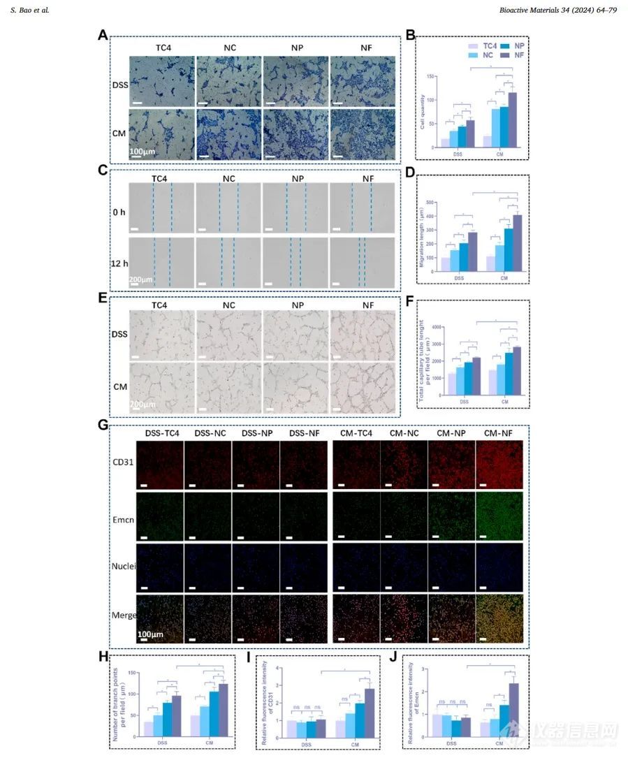
Figure 4. Angiogenesis of human umbilical vein endothelial cells (HUVECs) in direct stimulation sample (DSS) and conditioned medium (CM) modes respectively. (A) Crystal violet staining image of invaded HUVECs on the Transwell chamber membrane at 24 hours, (B) Count of invaded cells. (C) Scratch images at 0 and 12 hours, (D) Quantification of migration length of HUVECs. (E) Vascular lumen formation image of HUVECs, (F) total capillary length at 12 hours, (H) number of branch points per field of view. (G) Fluorescent staining images and relative intensities of CD31 (red) and endothelial mucin (Emcn, green) in HUVECs at 5 days. *p<0.05。
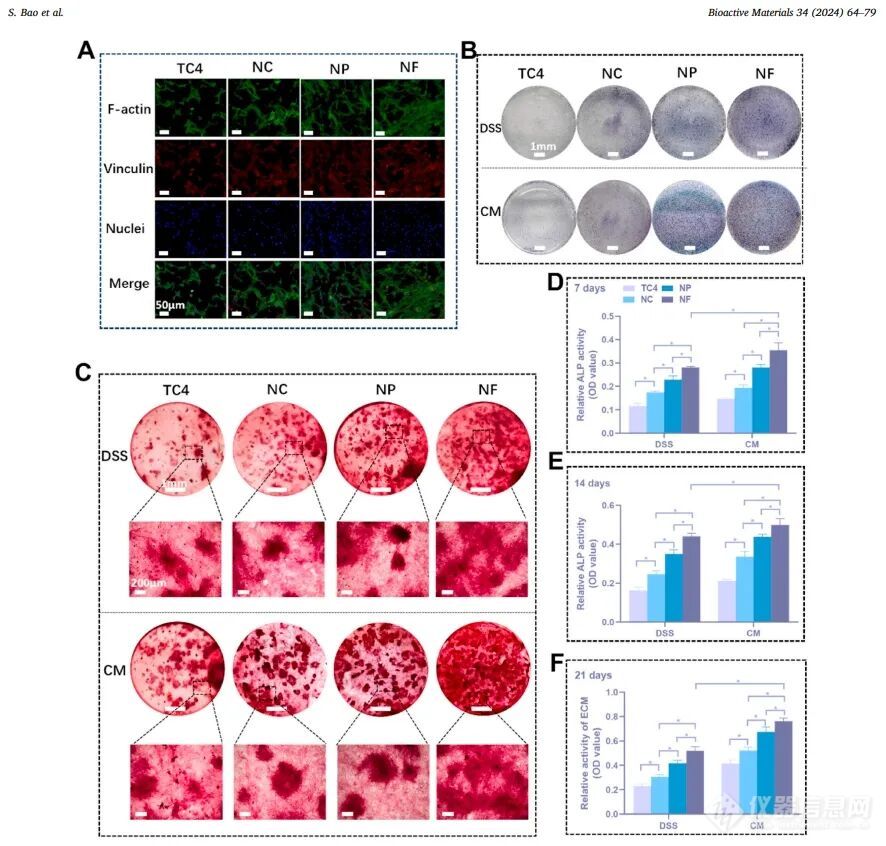
Figure 5. Adhesion, osteogenic differentiation and mineralization of MC3T3-E1 cells: (A) Fluorescent staining of F-actin (green), vinculin (red) and nuclei (blue) in MC3T3-E1 cells cultured in direct stimulation sample (DSS) mode for 3 days. (B) Representative images of alkaline phosphatase (ALP) staining of MC3T3-E1 cells at 14 days, (C) Alizarin red staining images of mineralized nodules of MC3T3-E1 cells co-cultured in DSS and conditioned medium (CM) for 21 days respectively. (D, E) Quantitative ALP activity at 7 and 14 days, (F) Quantitative analysis of mineralized nodules at 21 days. *p<0.05。
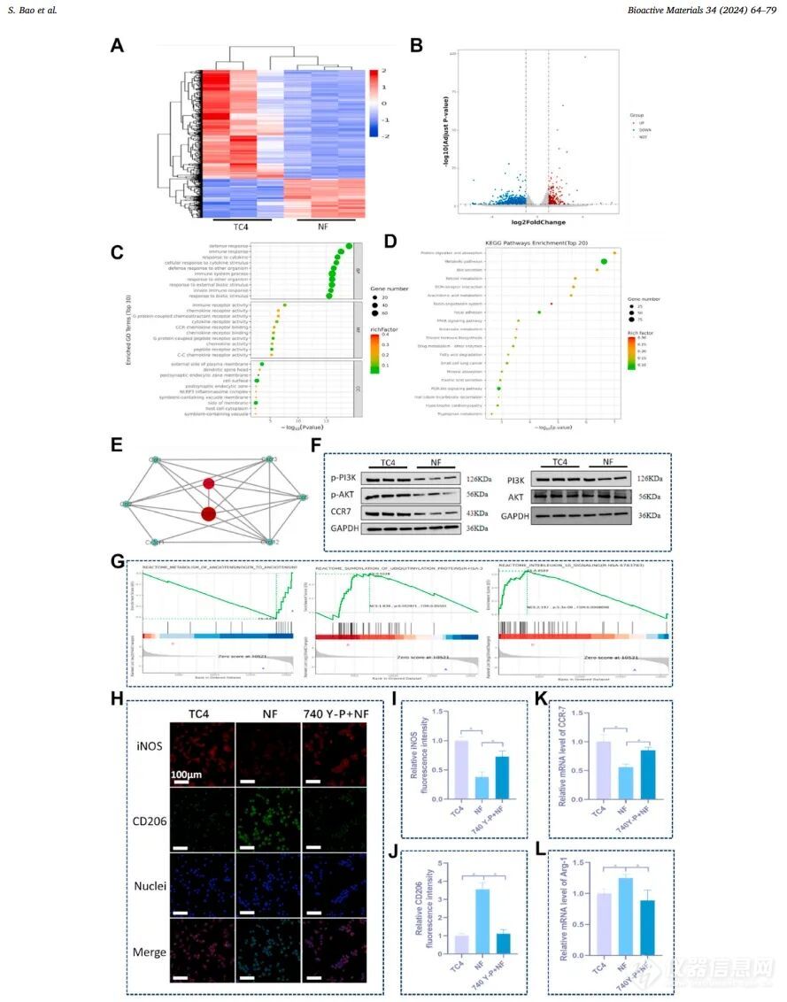
Figure 6. RNA sequencing analysis of RAW264.7 cells cultured on NF and TC4 as a control for 3 days, and verification of the inhibitory effect on inflammatory response. (A) Volcano plot, (B) heat map, (C) Gene Ontology (GO) pathway enrichment analysis of differentially expressed genes, (D) Kyoto Encyclopedia of Genes and Genomes (KEGG) enrichment analysis. (E) Protein-protein interaction (PPI) network analysis of differentially regulated pathways. (F) Representative Western blot of inflammation-related signaling pathways in RAW264.7 cells. (G) Enrichment analysis of regulatory gene pathways using KEGG and genomic databases. (H-J) Representative immunofluorescence images of inducible nitric oxide synthase (iNOS, red)/cluster of differentiation 206 (CD206, green) and their relative fluorescence intensity in RAW264.7 cells with and without 740Y-P treatment (NF and TC4 as controls), as well as (K) CCR-7 and (L) Arg-1 mRNA expression. *p<0.05。
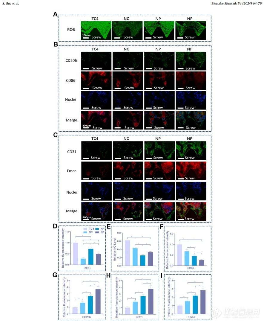
Figure 7. Fluorescent staining images of the tissue around rat femoral condyle screws: (A) Fluorescent staining images of reactive oxygen species (ROS) at 1 week after implantation; (B) Fluorescent staining images of CD86 (red), CD206 (green) and cell nuclei (blue) at 1 week after implantation; (C) Fluorescent staining images of CD31 (green), endothelial mucin (Emcn, red) and cell nuclei (blue) at 2 weeks after implantation. (D) Relative fluorescence intensity of ROS and (E) NO levels. Statistical relative fluorescence intensity of (F) CD86, (G) CD206, (H) CD31, and (I) Emcn. *p<0.05。
CT characterization results
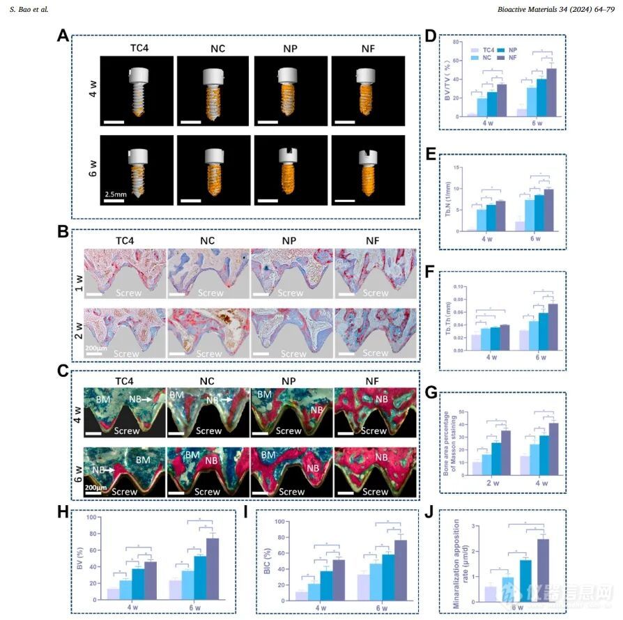
Figure 8. Rat femoral condyles implanted with bare and nanoOsseointegration status at 1, 2, 4 and 6 weeks after coating TC4 screws: (A) Micro-CT reconstructed image of newly formed bone (yellow) around the screw (white); (B) Masson stained image of new bone adjacent to the screw; (C) VanGieson (VG) stained image of new bone adjacent to the screw (NB: new bone; BM: bone marrow). (D) Quantitative ratio of new bone volume to total area around the reconstruction screw (BV/TV, %); (E) Trabecular bone number (Tb.N, 1/mm); (F) Trabecular bone thickness (Tb.Th, 1/mm) calculated from Micro-CT reconstructed images. (G) Masson stained bone area percentage. (H) Quantitative percentage of new bone volume (BV, %) and (I) bone-to-implant contact ratio (BIC, %) calculated from VG stained images. *p<0.05。
Quantitative analysis of new bone formation:The nanocerium oxide coating (especially the NF group) significantly promoted new bone formation at different time points after implantation, and its bone volume fraction (BV/TV), number of trabecular bone (Tb.N), and trabecular bone thickness (Tb.Th) were significantly higher than those of the control group TC4. For example, at 6 weeks after surgery, the BV/TV of the NF group reached approximately 80%, which was significantly better than that of other groups (Figure 8A-D).
Comparison of coating effects: The amount of new bone formation showed a trend of NF>NP>NC>TC4. The NF group had formed continuous mature bone 4 weeks after surgery, while the TC4 group only had a small amount of new bone (Figure 8C-H).
Vascularization support: NF coating provides a favorable vascular microenvironment for bone regeneration by promoting the formation of H-type blood vessels (CD31^hi/Emcn^hi) (Figure 7C-I).
Research conclusion
In this study, a nanomorphology (NC, NP and NF) ceria (CeO₂₋ₓ) coating with multiple free radical scavenging functions was prepared on the surface of titanium alloy (TC4) by hydrothermal method. Its function is equivalent to a nanozyme. Nano-CeO₂₋ₓ coating (especially NF) induces M2 polarized macrophages that are beneficial to bone immunomodulation and vascularized osseointegration by scavenging ROS and NO. In in vitro experiments, nano-CeO₂₋ₓ coatings (especially NF) showed obvious vascular regeneration and osteogenesis in DSS and CM modes. The CM mode showed a more significant enhancement effect due to the anti-inflammatory cytokines and growth factors secreted by M2 macrophages. The inflammation, angiogenesis and osteogenesis of the tissues around the nano-CeO₂₋ₓ-coated TC4 screws in the rat femoral condyle further confirmed that NF has osteoimmunomodulation and osseointegration functions. The underlying mechanism is that NF inhibits the macrophage PI3K-AKT signaling pathway, which plays a key role in macrophage polarization. In summary, the nano-CeO₂₋ₓ coating with the function of scavenging various free radicals contributes to immune regulation and subsequent osseointegration, providing promising ideas for the surface structure and chemical design of bone implants.
original document
[1]ShusenBao,DongmeiYu,ZhenTang,etal.Conformationallyregulated“nanozyme-like”ceriumoxidewithmultiplefreeradicalscavengingactivitiesforosteoimmunologymodulationandvascularizedosseointegration[J].BioactiveMaterials,2024,34:64-79.https://doi.org/10.1016/j.bioactmat.2023.12.006
Paper application equipment
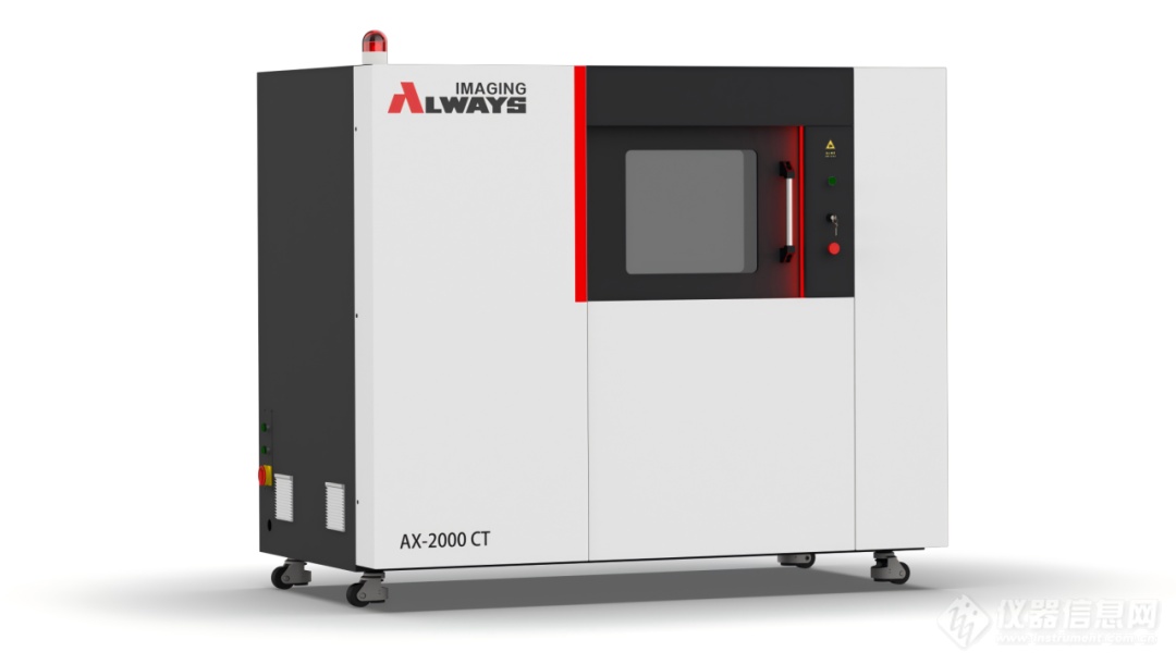
In this study, AX-2000CT played a key role. It can perform high-resolution scanning of titanium alloy samples modified with nano-cerium oxide coating on the surface, clearly showing the microstructure, thickness changes and bonding of the coating with the substrate.At the same time, for the titanium alloy screws and surrounding tissues after implantation in the rat femoral condyle, AX-2000CT can accurately capture the details of new bone formation at different time points, including information such as the growth, distribution and mineralization degree of bone trabeculae.Scanning these samples provides important visual data for studying the impact of nanocerium oxide coatings on osseointegration, which effectively promotes experimental progress.
thisIn addition, AX-2000CT is also suitable for non-destructive three-dimensional analysis of biomaterials such as bioceramics and polymer scaffolds and related tissue samples, providing scientific researchers with high-contrast microstructure visualization data. This technology provides a three-dimensional visualization basis for elucidating the interface effect of bone implant surface modification on bone tissue by accurately capturing key parameters such as material porosity and interface bonding characteristics.
View More from This Booth

The new version of the "Measures for the Classification of Radiographic Devices" is implemented: the CT classification and determination methods usher in important adjustments
Customer Literature | Research on micro-nano hemostatic materials based on beetle pulse jet bionic structure
Aoying Case | Digital model comparison (comparison between physical object and design model): efficient and excellent non-destructive inspection method for the quality of plastic (casting) parts

Aoying Science | The difference between DR and CT: the "fluoroscopy eye" and "tomography microscope" of non-destructive testing
Recommended Products

Always Imaging metrology microfocus industrial CT system AX-3000CT-M
US$ 489510.00-769231.00
Model:AX-3000CT-M